Dual waveform capnogram
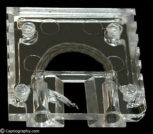
This capnogram occurs due to sampling leaks in the sampling tube cracks or a loose connection between the sampling tubes and the monitor. It has also been reported due to breaks in the water filter of the carbon dioxide analyzer.

(Provided by Simon Body and James Philips)
This shape of a capnogram can result in differential lung emptying of the lungs. For example, one may be a normal lung (transplanted lung) and the other a diseased lung. The normal lung has greater carbon dioxide to expire than the diseased lung.
In addition, a dual capnogram can result during endobronchial intubation and in patients with severe kyphoscoliosis.
Tripati et al. have described another capnogram with a “tail up,” as shown below, during the use of an accidentally crushed sampling tube with a slit-like hole.

The authors investigated the mechanics involved in 40 consenting ASA I patients undergoing general anesthesia. They observed the “tail up” capnogram with lower end-tidal CO2 values with a slit the sampling tubes as compared to the intact sampling tubes during IPPV. However, no such deformity was observed during spontaneous ventilation, although end-tidal CO2 was lower than normal.
A similar mechanism as described above may be operative during IPPV. However, during spontaneous ventilation, there is no positive pressure (in fact, there is slight negative pressure). Therefore, the dilution of sampling gases through the leak continues in both respiratory cycle phases. This gives a normal shape to the capnograms but with lower end-tidal CO2 values as compared to intact or undamaged sampling tubes.
Reference:
Tripati M, Pandey M. Atypical “tails up” capnogram due to breach in the sampling tube of side-stream capnometer. J Clin Monit 2000;16:17-20.

 Twitter
 Youtube


