Capnography in Veterinary Sedation
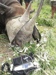
It has been recognized that monitoring respirations in patients during sedation for out of the operating room procedures is valuable in preventing the occurrence of hypoxic episodes. The value of monitoring respirations using capnography has migrated to veterinary anesthesia to protect lives of precious animals. The picture below demonstrates the use of capnography during sedation in Rhino in Africa. (courtesy: Robin W. Radcliffe, DVM, DACZM, Rhino Conservation Medicine Program, Adjunct Assistant Professor of Wildlife and Conservation Medicine International Rhino Foundation / Fossil Rim / Cornell University)

Morkel et al, measured acid-base balance and ventilation during sternal and lateral recumbancy in 36 field immobilized black Rhinoceros receiving oxygen insufflation. The animals were immobilized in Namibia from a helicopter, by remote intramuscular injection of etorphine HCl, azaperone, and hyaluronidase. They measured PETCO2, PVCO2, SPO2, HCO3 in lateral position and sternal position. For a similar PCO2, PETCO2 was higher in sternal position as compared to lateral recumbency position. The authors atrribute this to decreased physiological dead space as a result of better ventilation upon assuming sternal position from later recumbency. It would have been interesting to determine airway flows and ventilation in the two postions. Analysis of capnograms in the two positions would have also been useful. The additional information could determine if there was partial airway obstruction in the lateral position. Nonetheless, a notable feature of this study was that all animals were acedemic and hypoxic on capture. The information in this study will be very useful to improve the care of these animals upon capture.
Reference:
Morkel P.vdB, Radclifee RW, Jago M, Preez P du, et al. Journal of Wildlife Diseases 2010;46(1):236-45.

 Twitter
 Youtube


