Capnography as a non invasive-monitor of PaCO2 during thoracic anesthesia
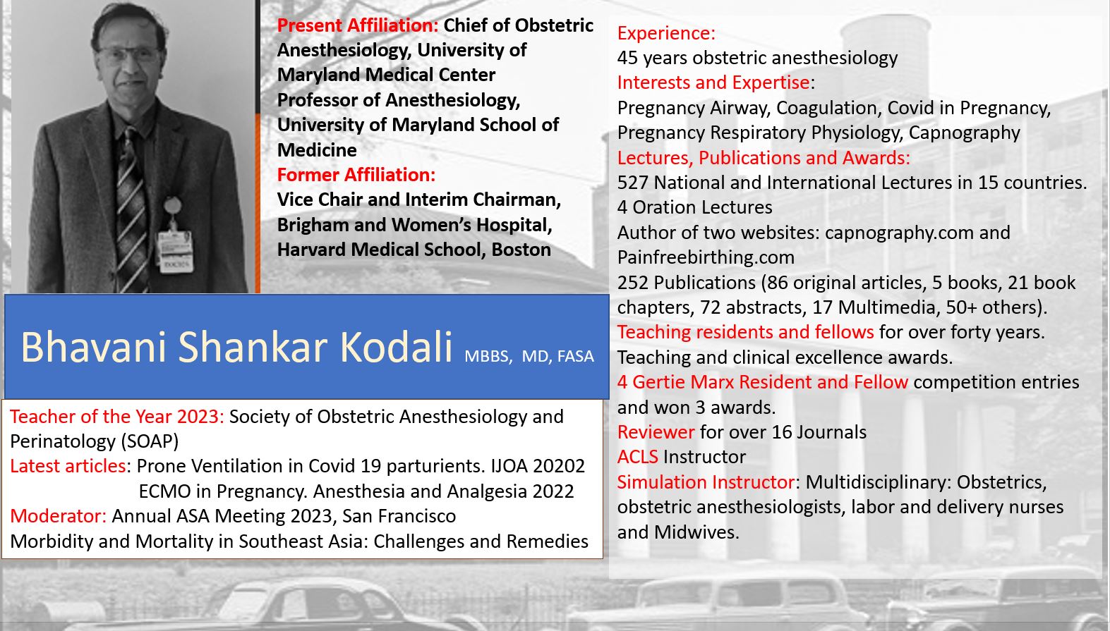
Bhavani Shankar Kodali MD
The arterial to end-tidal PCO2 difference is dependent on the underlying pulmonary disease. The most common reason for thoracotomy in adults is surgical treatment of lung cancer. Patients with lung cancer are almost exclusively smokers, and therefore have airways disease, and hence the PaCO2-PETCO2 gradient during anesthesia will be increased even in the supine position.1 In the 17 patients studied by Werner et al,2 mean PaCO2-PETCO2 from each lung was approximately 5 mm Hg in the supine position at a PaCO2 of 28 mm Hg and ventilatory frequency of 10/min. The lungs receive an approximately equal share of ventilation in the supine position, whereas, the ventilation of the upper lung is increased in the lateral position.2 However, the blood flow to the lungs is gravity dependent resulting in a decreased blood flow to the upper lung. This results in a decrease in the PETCO2 from the upper lung in the lateral position. Therefore, the PaCO2-PETCO2 as measured by Werner et al2 was zero for the lower lung and 11 mm Hg for the upper lung. Since the ventilation of the upper lung increases in the lateral position, it may surmised that if PETCO2 measured in the combined expirate of both lungs, the PaCO2-combined PETCO2 difference would be large.1,2
An incision of the chest wall produces an increase in mean pulmonary artery pressure2 which produces an increase in the CO2 elimination of the upper lung and a decrease in physiological dead space. The upper lung PaCO2-PETCO2 gradient is reduced when the mean pulmonary artery pressure increases as a result of surgical stimulation.
Furthermore, opening of the pleura increases CO2 elimination in the upper lung and thereby decreases PaCO2-PETCO2. Retraction of the lung produces exactly opposite effects. The lower lung gradient is not greatly effected with these maneuvers; it remains small. If combined end-tidal CO2 monitoring is performed, the PaCO2-PETCO2 would decrease when the pleura is opened, and increase again during retraction.
One lung ventilation:1,2
When the upper lung ventilation is discontinued to facilitate surgery, perfusion to the upper lung does not cease completely unless the pulmonary artery is clamped. In the absence of such clamping, there is a right to left shunt to the upper lung, the effect of which is to increase PaCO2, thus PaCO2-PETCO2 increases. However, this PaCO2-PETCO2 may not be any greater than the original combined two lung PaCO2-PETCO2.
Affect of prolonged expiratory maneuvers on PaCO2-PETCO2 during thoracotomy:3
In 16 patients undergoing thoracoabdominal esophagectomy, the affect of two prolonged expiration maneuvers to improve prediction of PaCO2 from PETCO2 were studied. PCO2 at the end of a simple prolonged expiration (PE1CO2), and PCO2 at the end of a prolonged expiration preceded by sustained hyperinflation of the lungs (PE2CO2), were measured during laparotomy, in the lateral thoracotomy position during two-lung ventilation, and after transition to one lung ventilation. PaCO2-PETCO2 was 9.75 (SD 3) mm Hg during laparotomy and this remained stable throughout the study. Both maneuvers decreased the mean arterial to peak expired PCO2 difference particularly during one lung ventilation. These results are in agreement with the results obtained by Bhavani-Shankar et al, where squeeze PETCO2 decreased PaCO2-PETCO2 in pregnant patients undergoing laparoscopic surgery.
Table from reference 3
| Measurement mean (SD) mm Hg | PaCO2-PETCO2 | PaCO2-PE1CO2 | PaCO2-PE2CO2 |
| End of abdominal procedure | 9.75 (3) | 6 (3.7) | 4.5 (3.7) |
| TLV for 20 min | 10.5 (3.7) | 7.5(4.5) | 3 (4.5) |
| OLV for 20 min | 9.75 (3) | 3 (3.7) | – 0.75 (3.7) |
| OLV for 50 min | 10.5 (3.7) | 2.2 (4.5) | -1.5 (3.7) |
| TLV after skin closure | 9 (3.7) | 3.7 (5.2) | 0.75 (4.5) |
However, the end-expiratory PCO2 obtained with each maneuver during laparotomy and thoracotomy agreed poorly with PaCO2. The authors3 suggest that these maneuvers should no longer be recommended to improve estimation of PaCO2 from PETCO2 during anesthesia.
Arterial to end-tidal CO2 difference after bilateral lung transplantation.
| Variable | Time after lung transplantation |
| Mean (SD) | 10 min | 1 hr | 3 hr | 12 hr | 24 hr |
| PaCO2-PETCO2 mm Hg | 16 (5) | 14 (5) | 9 (4) | 6 (3) | 5 (3) |
| (PaCO2-PETCO2)/PaCO2 | 0.36 (0.13) | 0.33 (0.13) | 0.21 (0.12) | 0.15 (0.07) | 0.11 (0.06) |
The time course of the arterial to end-tidal PCO2 difference suggests a rapid improvement of this post-transplant ventilation/perfusion mismatch. After 24 hrs, the values were close to the physiological range, which is supposed to be 4-5 mm Hg. A possible explanation of these findings could be ischemia-reperfusion injury that affects microcirculation. An impaired distribution of pulmonary blood flow with unperfused alveoli would clinically appear as alveolar dead space as is seen immediately following lung transplantation. The ventilation/perfusion mismatch normalized in about 24 hrs indicating a redistribution of pulmonary blood flow and recovery of microcirculation.
Arterial to end-tidal carbon dioxide difference during anesthesia for thoracoscopy:
Presently, thoracoscopy is the procedure of choice in most patients requiring thoracic surgery, especially those patients with severe underlying lung disease.6,7 Srinivasa et al7 studied the difference between PaCO2 and PETCO2 during thoracoscopic surgery in ten patients scheduled for elective thoracoscopic procedures. All patients had general anesthesia induced by IV propofol, fentanyl and vecuronium. A double lumen endo-tracheal tube was positioned with the aid of a fiber optic bronchoscope. Anesthesia was maintained with 100% oxygen, desflurane, fentanyl and vecuronium. Ventilation was kept at, tidal volume (TV) 10 ml/kg, respiratory rate (RR) of 10 bpm and an I:E ratio of 1:2 while on two lung ventilation. TV was 7 ml/kg, RR of 10 bpm and an I:E ratio of 1:2 while on one lung ventilation (OLV). The FiO2 during all measurements was kept at 1.0. Arterial blood gas was sampled 10 min after the patient was in lateral decubitus position while on two-lung ventilation (T1). The second sample (T2) was10 min after the introduction of trocars (OLV). The last sample (T3) was taken 10 min after restarting two-lung ventilation.
Results: The mean FVC was 2.6 ± 0.8 L (79 ± 19% predicted) and the FEV1 was 1.9 ± 0.7 L (76 ± 26% predicted). Table1 shows demographic data of the patients. The mean PaCO2 to PETCO2 difference at times T1, T2 and T3 were 5.5 ± 4, 7.4 ± 5, 6.8 ± 4 mm Hg respectively. The lowest value noted for PETCO2 was 27 mm Hg and the highest value for PaCO2 was 52 mm Hg during the study (Fig.2). The difference between the PETCO2 and PaCO2 during the various time intervals was not statistically significant.
| Time | PETCO2 | PaCO2 | Difference |
| T1 | 32 ± 3 | 38 ± 5 | 5.5 ± 4 |
| T2 | 35 ± 4 | 42 ± 5 | 7.4 ± 5 |
| T2 | 32 ± 3 | 39 ± 6 | 6.8 ± |
References:
(1). Fletcher R: The Arterio-End-Tidal CO2 difference during cardiothoracic surgery. J. cardiothorac. Anesthesiol. 4:105-117, 1990.
(2). Werner O. Malmkvist G, Beckman A, et al: CO2 elimination from each lung during endobronchial anaesthesia. Br. J. Anaesth. 56: 995-1001, 1984.
(3). Tavernier B, Rey D, Thevenin J, Triboulet P, Scherpereel P. Can prolonged expiration manoeuvres improve the prediction of arterial PCO2 from end-tidal PCO2? Brit J Anaesth 1997;78:536-40.
(4). Bhavani Shankar K, Steinbrook R, Mushlin PS, Freiberger D. Transcutaneous carbon dioxide monitoring during laparoscopic surgery in pregnancy. Canadian J Anaesth 1998;45:164-9.
(5) Jellinek H, Hiesmayr M, Simon P, Klepetko W, Haider W. Arterial to end-tidal CO2CO2 tension difference after bilateral lung transplantation. Crit Care Med 1993;21:1035-40.
(6) Horvath KA: Thoracoscopic transmyocardial laser revascularization. Ann.Thorac.Surg. 1998; 65: 1439-41.
7) Kotloff RM, Tino G, Bavaria JE, Palevsky HI, Hansen-Flaschen J, Wahl PM, Kaiser LR: Bilateral lung volume reduction surgery for advanced emphysema. A comparison of median sternotomy and thoracoscopic approaches. Chest 1996; 110: 1399-406.
(8) Srinivasa V, Kodali BS, Bean T, Hartigan PM. Arterial to end-tidal carbon dioxide difference during thoracoscopic surgery. Anesthesiology ASA abstracts 2004;A1556.

 Twitter
 Youtube


