Terminology of Capnograms
Over the last two decades, time capnography has become a standard of monitoring in anesthesia practice in many countries. Along with the acceptance of this technology, however, there has also been a considerable proliferation of terminology representing the various components of a time capnogram. This ambiguity in terminology has been a source of confusion to readers.
Past Terminology
 |
 |
 |
 |
For instance, numerous terms, such as PQRS, ABCDE, EFGHIJ, and phases I through IV, have been used to depict the various components of a time capnogram.1-12 Some have used “phase IV” to designate the terminal upswing at the end of phase III, which is occasionally observed in capnograms recorded in pregnant and obese subjects. Others have used “phase IV” to designate the descending limb of a time capnogram. In much the same way that the nomenclature of the various segments of the ECG have been standardized, it is necessary to define and standardize the nomenclature used to designate the various components of a time capnogram.
A standard terminology facilitates teaching, comprehension, communication, and research A terminology representing various phases of a time capnogram, and based on logic, convention, and tradition, has been described by Bhavani-Shankar et al several years ago.2 This terminology is currently being adapted by several authorities including Nunn’s Respiratory Physiology.13 The logical and conventional basis of this terminology is summarized as follows.23
Current Terminology

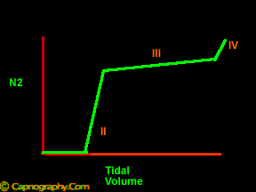
In 1949, Fowler described SBT-N2 (single-breath test for nitrogen) to study uneven ventilation in lungs where instantaneous nitrogen concentrations are plotted against expired volume.14 The resulting nitrogen curve is divided into four phases: phase I, phase II, phase III, and phase IV. When the instantaneous CO2CO2 concentration is plotted against expired volume , the resulting curve resembles an SBT-N2 curve in shape and is called an SBT-CO2 curve. An SBT CO2 curve is also traditionally divided into three phases: I, II, and III, and, occasionally, a phase IV, if present.1,2,15. Phase IV does not occur normally, but may be seen under certain circumstances, as described in the physiology section. The physiologic mechanism responsible for phases I, II, and III is similar in SBT-N2 as well as in SBT-CO2 curve. However, the mechanism resulting in phase IV in SBT-CO2 may be different from that in an SBT-CO2 curve, as explained in the physiology section.
 |
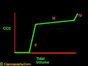 |
Unlike an SBT-CO2 trace, a time capnogram has an inspiratory segment in addition to the expiratory segment. There is no inspiratory segment in an SBT-CO2 curve, as, by definition, a SBT-CO2 trace is a plot of PCO2 and expired volume. However, the expiratory segment of a time capnogram resembles an SBT-N2 curve and an SBT-CO2 curve in shape. Furthermore, the physiologic mechanism responsible for the shape of the expiratory segment is similar to that in either SBT-N2 curve, or SBT-CO2 curve.2 h4: Hence, it is prudent conventionally and logically to also consider the expiratory segment of time capnogram as three phases: I, II, and III as in SBT-N2/SBT-CO2 curve. Occasionally, at the end of phase III, a terminal upswing (phase IV) seen in an SBT-CO2 curve or an SBT-N2 curve, may occur in a time capnogram. The details of phase IV are discussed in the physiology section :
 |
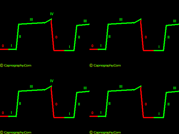 |
Current terminology is summarized as follows.
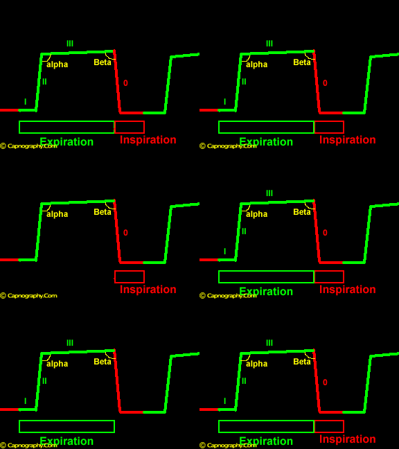
A time capnogram can be divided into inspiratory (phase 0) and expiratory segments. The expiratory segment, similar to a single breath nitrogen curve or single breath CO2 curve, is divided into phases I, II and III, and occasionally, phase IV, which represents the terminal rise in CO2 concentration. The angle between phase II and phase III is the alpha angle. The nearly 90 degree angle between phase III and the descending limb is the beta angle.
Limitation of time capnogram
The assumption that expiration ends at the commencement of down-stroke is not necessarily true all the time. In a time capnogram, the beginning and the end of an inspiratory segment, and the beginning and the end of expiration (expiratory time) cannot be delineated accurately without superimposing the simultaneously recorded respiratory flows. Expiration begins somewhere in the horizontal line before the actual upstroke, and ends somewhere on the phase III, reminder of phase III being expiratory pause. For further understanding of this concept, please refer to the references 2 and 3 below, and the ‘Capno-pitfalls’ section of this website :
Reference
*1. Bhavani-Shankar K, Moseley H, Kumar AY, Delph Y. Capnometery and Anaesthesia: Review article. Can J Anaesth 1992;39:6:617-32.
* 2. Bhavani-Shankar K, Kumar AY, Moseley HSL, Ahyee-Hallsworth R. Terminology and the current limitations of time capnography: A brief review. J Clin Monit 1995;11:175-82.
* 3. Fletcher R, Jonson B. Deadspace and the single breath test for carbon dioxide during anesthesia and artificial ventilation. Br J Anaesth 1984;56:109-19.
* 4. Moon RE, Camporesi EM. Respiratory monitoring. In Miller RD, ed. Anesthesia, ed 5. New York: Churchill Livingstone, 2000:1255-95.
* 5. Good ML, Gravenstein N. Capnography. In Ehrenwerth J, Eisenkraft JB, eds. Anesthesia Equipment, ed 1. Boston: Mosby, 1993:237-48.
* 6. Hardwick M, Hutton P. Capnography: Fundamentals of current clinical practice. Curr Anaesth Crit Care 1990;3:176-80.
* 7. Adams AP. Capnography and pulse oximetry. In: Atkins RS, Adams AP, eds. Recent advances in anaesthesia and intensive care. London: Churchill Livingston, 1989:155-175.
* 8. Sweadlow DB. Capnometry and capnography: The anesthesia disaster early warning system. Seminars in Anesthesia 1986;3:194-205.
* 9. Ward SA. The capnogram: Scope and limitations. Sem Anesth 1987;3:217:28.
* 10. Curley MAQ, Thompson JE. End-tidal CO2 monitoring in critically ill infants and children. Pediatr Nurs 1990;16:397-403.
* 11. Kalenda Z. Mastering infrared capnography. Utrecht, The Netherlands: Kerckebosch-Zeist, 1989:p101.
* 12. Bhavani-Shankar K, Philip JH. Defining segments and phases of a time capnogram. Anesthesia and Analgesia 2000;(4):973-7.
* 13. Nunn’s Appplied Respiratory Physiology. 5th edition. Boston: Butterworth-Heinemann, 2000;243
* 14. Fowler WS. Lung function studies V. Respiratory dead space in old age and in pulmonary emphysema. J Clin Invest 1950;29:1439-44.
* 15. Fletcher R. The single breath test for carbon dioxide (Thesis). Lund, Sweden, 1980.

 Twitter
 Youtube


